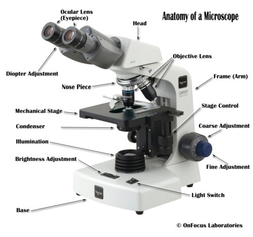
The Compound Light Microscope
Helpful Links
Microscope Key Points, Diagrams, and Quizzes
Exploring Magnification: Power of Ten video
How to use the microscope video
Appendix A of your Lab Manual – Use and Care of Microscopes
Reminders
- Always start with the 4X objective lens. Find the specimen and focus using the coarse focus, then the fine focus knob. Then progress to the 10X objective lens, then 40X lens and oil/100X lenses. Be sure to focus at EACH new objective lens using ONLY the fine focus knob. Do not skip using a lens. Always progress one lens at a time.
- Label all drawings with date, time, place and any relevant notes.
- Record all observations; don’t rely on memory.
- Make sure to record what you SEE, not what you THINK you should see.
- Detail is key, pay attention to even the smallest things.
- Include an indication of size (total magnification, field of view, size bar or lens used in relation to the field of view)
In lab you will use prepared slides to practice using a compound microscope – draw what you see and label at least 2 structures you can see within your organism.
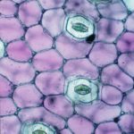
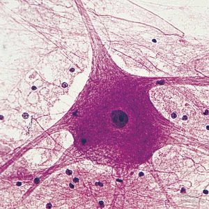
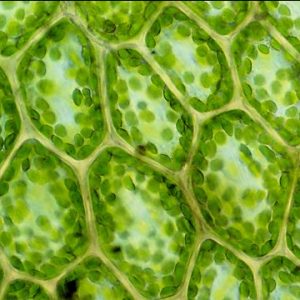
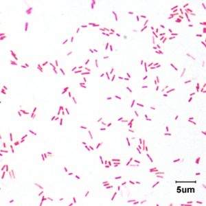
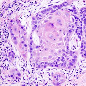
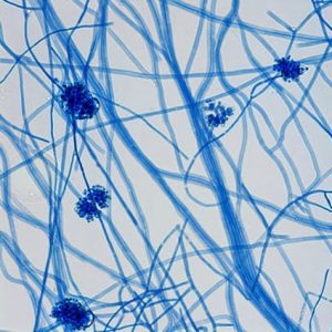
- Below are images of bacteria, animal, plant and fungal cells. Can you tell which are animal? Which are plant? Be able to defend your answer. What additional information would be helpful in making your decision?
1. 2.
2.  3.
3. 
4. 5.
5. 6.
6.
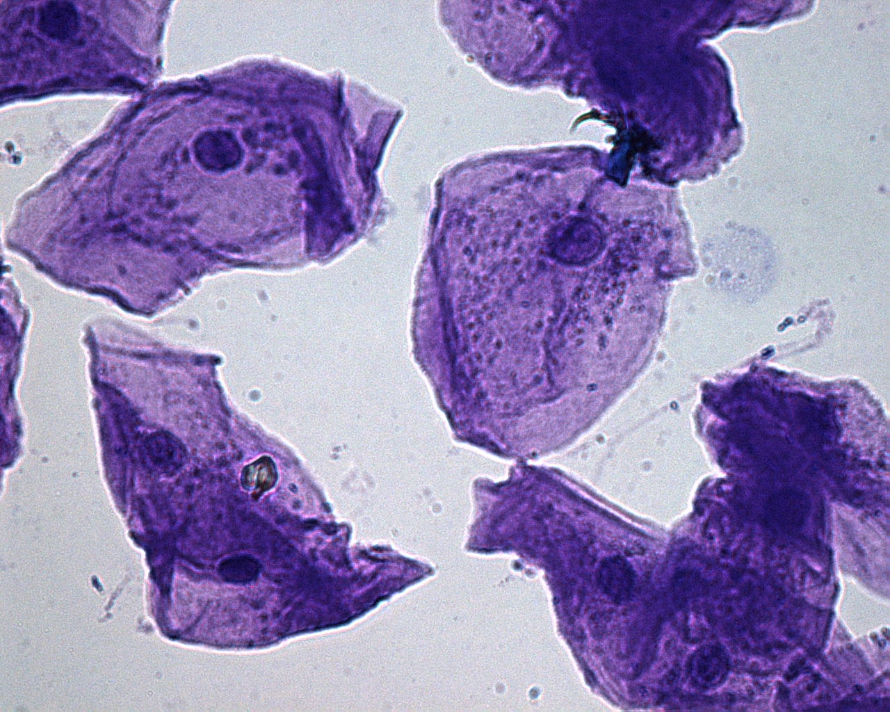
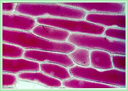
Simple Staining with Animal and Plant Cells (Eukaryotic Cells)
Simple staining is the addition of ONE type to stain that will allow you to better see cells and/or structures under the light microscope.
In lab you will view and draw cheek and onions cells that were stained with toluidine blue using your compound microscope.
How would you describe the animal cells? The plant cells? Size, shape organization? What structures and organelles can you see that help you identify the type/kingdom of these cells? What cellular organelles can’t you see? Why can’t you see them?
Video: Osmosis in Red Onion Epidermis – (1:41) flushing various concentrations of solution through these plant epidermal cells.
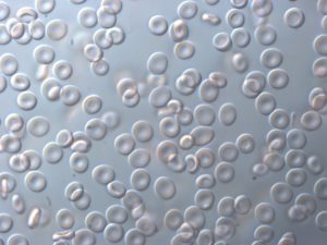
Video: Osmosis in Red Blood Cells-(3:47) see how red blood cells appear in isotonic, hypertonic and hypotonic soloutions.
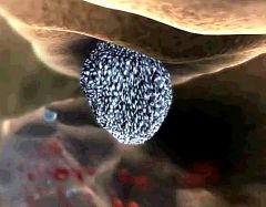
Video: Cellular Visions: Inner Life of the Cell video (3:12)- Read the summary below before you watch the video. Click on the link and scroll down and click the play video arrow.
Video Background: This video starts with a view of the inside of a blood vessel. You will see the red blood cells rushing past and the much larger white blood cells rolling along the wall of the blood vessel. The white blood cell receives an extracellular signal and the majority of the video shows what happens inside the white blood cell as it responds to that signal. The final scene shows the white blood cell reshaping (due to the conformational changes of the cytoskeleton) and slipping through the endothelial cells of the blood vessel wall to the site of inflammation.
As you watch the video, how many structures/organelles can you see? Can you find -protein filaments, lipid rafts, plasmamembrane, actin filaments, microtubules, motor proteins, transport vesicles, mitochondria, centrosomes, nucleus, nuclear pore, mRNA, ribosomes making proteins, ER, golgi apparatus, vesicles transported back to the plasmamembrane surface.
Article: NCSU Researcher “Stopping Cancer in its Track” – Examines the role to which the cytoskeleton/actin is involved in movement of cancer cells.
[callout heading=”Assignment: Cells” headingicon=”noicon” textalign=”textcenter” url=”https://wordpress-projects.wolfware.ncsu.edu/bio-183-lab-yhnn8gz/wp-content/uploads/sites/74/2020/02/CellUnitAssignS20.pdf” target=”true” type=”basic” bgcolor=”huntyellow”]This assignment is worth 15 points. It is due at the start of lab next week (Feb. 17-21).</p>
<p>Click on this box to download the assignment.[/callout]