Part I: Policies and Procedures
Read the first pages of the lab manual on policies and procedures (p.v. -ix) and sign page ix for turning in to your lab instructor. If you have any questions, please let your lab instructor know during the second week of labs.
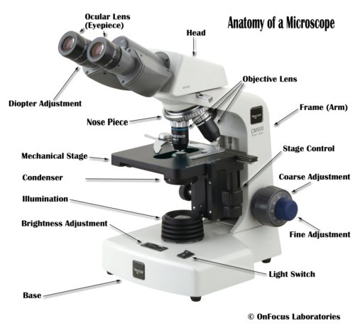
Part II: Learning how to use the microscope
See the video below for the basics on microscope use
Part III: Microscope Calculations – Total Magnification, Field of View, and Cell Size
a. Go to page 3 of the lab manual and complete Table 1-1. Total magnification for each objective
b. Calulating field of view and cell size
Read pages 4 and 5 of the lab manual and complete table 1-2. See the video below for a demonstration of how this is done.
Part IV: Examining Live Specimens:
Click on the microscope simulation below and follow the directions. Start with either a plant cell or animal tissue, then make sure to do both.
The power of making careful observations, read p. 5 in the lab manual.
- Observations: Statements determined by using any one of a person’s five senses (empirical reasoning). It is typically the first step in the Scientific Method and can lead to hypotheses.
- We will be using Microscopes to make Observations of small living specimen and writing descriptions to practice Scientific Communication skills.
Reminders:
- Label all notes with date, time, and place.
- Record all results; don’t rely on memory.
- Make sure to record what you SEE not what you THINK should be there.
- Detail is key, pay attention to even the smallest things.
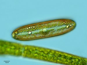
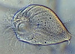
[callout heading=”ACTIVITY” headingicon=”noicon” textalign=”textleft” type=”basic” bgcolor=”bioindigo”]Choose two of the following specimens from the list below. Make observations and record them on page 7 of the lab manual. Your observations should be specific enough that anyone reading them would be able to identify the organism that you are describing. Assume that the total magnification is 100X and that the field of view is 1.04mm. Fill in the information (labels) on page 7 and draw your specimen. Do the same for specimen two using the back of page 7. Bring completed work to lab next time along with your filled in tables 1 and 2.[/callout]
Blepharisma
Stentor
Phacus
Rotifer
Euglena
[callout heading=”HOMEWORK: Library Tutorial and Assignment” headingtype=”p” headingicon=”noicon” textalign=”textcenter” url=”https://wordpress-projects.wolfware.ncsu.edu/bio-181l-zchxzbn/laboratory-units/laboratory-1/biology-181-library-tutorial/” type=”basic” bgcolor=”huntyellow”]Click this box to view a tutorial about scientific literature and how to find references to help Interpret/explain scientific observations and data from your experiments.[/callout]
Additional Resources:
The Compound Light Microscope
- Care and Use of Microscopes – See Appendix A in your Lab Manual
- Video on How to Use a Compound Microscope
- Directions for determing Total Magnification, Field of View Measurements and Cell Size can be found in your lab manual Unit 1 Activities One to Four.
- Basic Concepts of Microscopy
- Exploring Magnification: Power of Ten video (from 10 million light years away from the Milky Way down to 100 attometers !)