Scroll below for In-Person Lab
Introduction to the lab:
- Read the background material for this lab (pp. 149-152). After reading the background material, organize the indicated terms listed in the assignment sheet in a table, concept map, or any other representation that will help you remember and understand them.
- If making a table, explain each term.
- Want to know about concept mapping? Below are some resources:
How to make a concept map - Example concept map
- Free Concept Mapping Application
- Want to know about concept mapping? Below are some resources:
Activity One: Paramecium feeding (online videos) – See materials below
- Refer to pp. 153 & 154. Use the online videos on the lab website to learn about how the Paramecium feeds, type of digestion, and structures involved. The videos will substitute steps 1-4 & 6-7 of this activity.
- Use what you have learned to complete steps 5 & 8 in your lab manual.
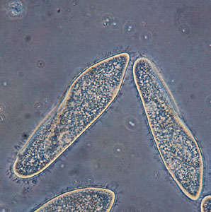
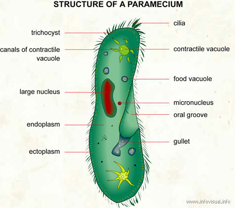
COMPOUND LIGHT MICROSCOPE (AT 400X) AND DIAGRAM (LEFT AND RIGHT IMAGE RESPECTIVELY)
- Description: ciliate Protozoan
- Habitat: stagnant freshwater
- Movement: cilia
- Nutrition: An oral groove is used to ingest nutrients and food vacuoles are responsible for digestion.
- Distinct Layers: The cytoplasm of protozoans can be divided into 2 layers. The ectoplasm is the more clear outermost layer and the endoplasm is the central area surrounding all organelles.
Paramecium:
- Description: ciliate Protozoan
- Habitat: stagnant freshwater
- Movement: cilia
- Nutrition: An oral groove is used to ingest nutrients and food vacuoles are responsible for digestion.
- Distinct Layers: The cytoplasm of protozoans can be divided into 2 layers. The ectoplasm is the more clear outermost layer and the endoplasm is the central area surrounding all organelles.
VIDEOS OF PARAMECIUM FEEDING (#1 IS WHAT YOU WOULD HAVE SEEN IN LAB)
- Paramecium eating Congo Red stained yeast cells. As the yeast cells enter the Paramecium, they are encapsulated into food vacuoles. When the yeast cells get digested by acidic enzymes, the red stain will change the yeast cells from red to blue.
- At higher magnification you can see the movement of the cilia around the perimeter of the cell used for movement, but also along the oral grove into the gullet.
- Closer still you can observe the cilia along the oral grove and the movement of food particles flowing into the cell. A food vacuole forms as particles get encapsulated to form a vacuole, which are then is released into the cytoplasm of the cell.
- After observing this feeding behavior, what terms can you apply to this type of feeding (Food Acquisition) and Digestion and Absorption of nutrients?
Activity Two: Hydra feeding (online videos) – See materials below
- Refer to pp. 154 & 155. Use the online videos on the lab website to learn about how the Hydra feeds, type of digestion, and structures involved. The videos will substitute Steps 1-3.
- Use what you have learned to complete Step 4.
- What key characteristics can you see above that helps you know that Hydra are part of the phylum Cnidaria?
- How do Hydra get food, ingest it, digest it, get the nutrients, and get rid of waste? -use the proper terms for these processes.
- Watch as a tiny Hydra rips its mouth open to feed. Here is a short article that further explains how this works).
VIDEOS OF HYDRA FEEDING (#2 IS SIMILAR TO WHAT YOU WOULD HAVE SEEN IN LAB FEEDING THE HYDRA DAPHNIA OR BRINE SHRIMP)
- Video of Hydra eating a mosquito larva. Note the overall structure of the Hydra and the smaller bulb-like stinging cells (nematocysts) along the tentacles. Though the mosquito larva is quite large, the lack of a skeleton and jelly like body allows the Hydra a lot of stretching to fit food items through the mouth and into the gastrovascular cavity for digestion. How does the waste then get out?
- Green Hydra eating 3 water fleas (Cyclops). The Green Hydra have green algal endosymbionts that provide some carbohydrates to the Hydra, but not so much that they still need to eat small zooplankton.
- Describe how Hydra get food, ingest it, digest it, get the nutrients, and get rid of waste?
- After observing this feeding behavior, what terms can you apply to to the Hydra in for feeding (Food Acquisition) and Digestion and Absorption of nutrients?
Activity Three: Bullfrog Dissection (p. 155-157)
In-Lab Dissection:
- Groups will work as a table to dissect a bullfrog. Follow the direction in your lab manual (step #1 on page 155). Examine the external of your frog. Can you find the indicated external structures? Are there any clues that let you know if this is a male or female frog?
- Work through the rest of the directions (steps 2-11). Answer all the questions and make the provided sketches and/or take appropriate pictures to label the indicated structures.
- If you take your own pictures of your dissection, you will need to label the main structures digitally. Scan/take pictures of your work from your lab manual and upload to Moodle with your answers to the end of Lab questions (see #6 below).
- Additional images and videos are provided below that may be helpful for drawing some of the anatomy and/or answering questions.
- For your Post-Lab Assignment – Answer questions #1-6 on pp. 165-166 of your lab manual. The questions encompass Activities 1-4. Do not copy and paste the information found in the lab manual or website. Use your own words and interpretations.
If you prefer not to participate in the Dissection:
- Use the provided images found in the Frog Dissection (remote) PPT presentation to complete the material covered on pages 155-157. These are nice clean images of a frog dissection.
- You will need to select View –> Slideshow. Then click through different regions of the frog and organ systems to view all the indicated structures in your lab manual.
- Additional images and videos are provided below that may be helpful for drawing some of the anatomy and/or answering questions.
- Make sure to fill in the material requested on p. 155-157 and complete Steps 7-11 in your lab manual.
- Draw/sketch the internal anatomy from the online frog dissection and label all organs in correct location. Do not take pictures of the online images to label. Scan/take pictures of your work from your lab manual and upload to Moodle with your answers to the end of Lab questions (#6 below). Please check to make sure your writing is legible.
- For your Post-Lab Assignment – Answer the end of lab Questions on pp. 165-166 of your lab manual. The questions encompass Activities 1-4. Do not copy and paste the information found in the lab manual or website. Use your own words and interpretations.
Images of Frog Dissections
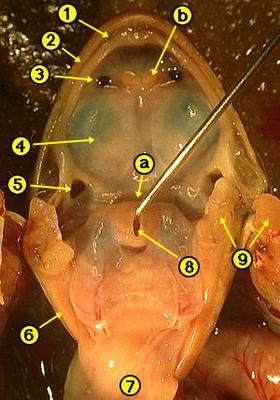 | Frog Oral Cavity
a. Esophagus opening b. Vomerine teeth 1. Groove of the maxilla 2. Maxillary teeth 3. Internal nare 4. Eye ball 5. Eustachian tube opening 6. Jaw ridge 7. Underside of the tongue 8. Glottis 9. Cut muscle and bone
|
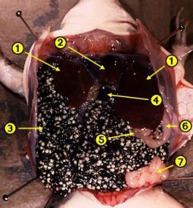 | Internal – Female Frog
1. Liver lobes 2. Heart 3. Ovary with eggs 4. Gall bladder 5. Small intestine 6. Stomach 7. Oviduct
|
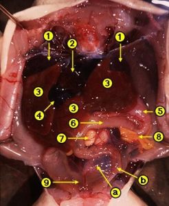 | Internal – Male Frog
a. Large intestine b. Small intestine 1. Lung lobes 2. Heart 3. Liver lobes 4. Gall bladder 5. Stomach 6. Small intestine 7. Testis 8. Fat Body 9. Urinary bladder |
Videos of a preserved bullfrog dissection
- Review of external anatomy and internal mouth structures. This video nicely shows the interesting attachment of the tongue and its function in capturing prey. You will also see the 2 types of teeth used to help hold prey: the maxillary and vomerine teeth.
- Review of Internal anatomy of the digestive, and the circulatory, respiratory and excretory systems.
Good written summaries and pictures of important frog structures and dissection. The discussion of the mouth structures explain how the tongue is used to capture prey and points out the 2 types of teeth (maxillary and vomerine teeth).
Activity Four: Mammals’ Teeth and Bird Bills (p. 158-166)
Part I: Mammals’ Teeth
- Read the background material and materials below, pp. 158-160.
- You will complete Tables 9-1 and 9-2 under the guidance of your lab instructor.
- In your lab groups, complete Tables 9-3 and 9-4. You will be examining the mammalian skulls with your group members.
Cool skull website – California Academy of Sciences – click on “Enter the 3D gallery”
Skull Structures and Muscles of Mastication – Additional Information to help interpret feeding habit clues seen in skulls beyond looking at teeth.
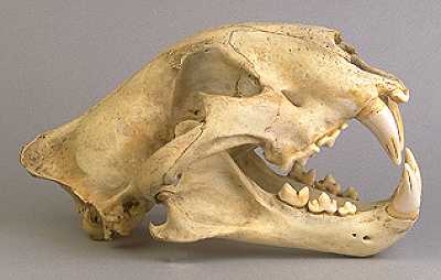
View Mammal Skulls
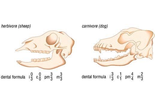
- For each skull you have examined, classify it as an herbivore, an omnivore, or a carnivore. For each, provide at least two justifications for your choice (for example, explain the type of teeth, eye placement, muscle attachments) and try to identify the type of animal.
- Based on your observations, which animal appears to possess the most heterodont dentition?
- Read the background material, p. 163 and review Bird Beaks and Bills below.
- In your lab groups, complete Table 9-5. You will be examining bird skulls and bills with your group members.
- For the bills provided, do you think that there might be another purpose other than diet for bill shape or color (defense, courtship display, etc.)? If yes, please explain.
- Using the sheet provided below, Bird Beaks and Bills, identify at least three bird species (other than those you have already examined) and describe how their bill is designed for acquisition of food. If you do not know many species of birds, you can do a quick websearch of bird images from Audubonorg. You may want to think of common backyard birds in your local region.
Part II: Birds’ Bills
View Bird Skulls
- Read the background material, p. 163 and review Bird Beaks and Bills below.
- In your lab groups, complete Table 9-5. You will be examining bird skulls and bills with your group members.
a. For the bills provided, do you think that there might be another purpose other than diet for bill shape or color (defense, courtship display, etc.)? If yes, please explain.
b. Using the sheet provided below, Bird Beaks and Bills, identify at least three bird species (other than those you have already examined) and describe how their bill is designed for acquisition of food. If you do not know many species of birds, you can do a quick websearch of bird images from Audubon.org You may want to think of common backyard birds in your local region.
Bird Beaks and Bills – Explore the various types of beaks and bills. Birds have various bill/beak sizes and shapes for a variety of reasons. However, the most important factor in determining beak shape and size is feeding habits.
What Big Teeth You Have – NCSU researchers looking at tooth root surface area can help determine the size of primates.
Activity Three: Bullfrog Dissection – mentioned earlier with the In-Lab Frog Dissection information.
- For your Post-Lab Assignment – Answer the end of lab Questions #1-6 on pp. 165-166 of your lab manual. These questions encompass Activites 1-4. Do not copy and paste the information found in the lab manual or website. Use your own words and interpretations.
- Draw, sketch or take pictures (if you did the In Person dissection) of the dissection and label all organs in the correct location. If you took your own pictures of the dissection, you will need to label the main structures digitally. Scan/take pictures of your work from your lab manual and upload to Moodle with your answers to the end of Lab questions (#1 above). Please check to be sure your writing is legible.
Gut Feelings Takes on New Meaning – Touch sensors in the gut lining respond to mechanical pressure and play an important role in digestion