Description and Size
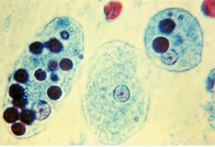
Entamoeba histolytica
This is a protist that infects predominantly humans and other primates. Diverse mammals such as dogs and cats can become infected but usually do not shed cysts (the environmental survival form of the organism) with their feces, so do not contribute significantly to transmission. The active (trophozoite) stage exists only in the host and in fresh feces; cysts survive outside the host in water and soils and on foods, especially under moist conditions on the latter. When swallowed they cause infections by excysting (to the trophozoite stage) in the digestive tract.
The cysts of Entamoeba histolytica, shown below, measure between 10 and 20 µm in diameter. The key identification feature is the number of nuclei; cyst will have 1 to 4 nuclei. In addition, the peripheral chromatin is generally evenly distributed. In the images below, two nuclei are clearly visible. Image on the left is iron-hematoxylin stain (1000x), and the image on the right is an iodine wet mount (1000x).

Amebiasis – Amebiasis is transmitted by fecal contamination of drinking water and foods, but also by direct contact with dirty hands or objects as well as by sexual contact. The infection is “not uncommon” in the tropics and arctics, but also in crowded situations of poor hygiene in temperate-zone urban environments. Individuals with a damaged or undeveloped immunity may suffer more severe forms of the disease. Human cases are diagnosed by finding cysts shed with the stool; various flotation or sedimentation procedures have been developed to recover the cysts from fecal matter.
Infections that sometimes last for years may be accompanied by 1) no symptoms, 2) vague gastrointestinal distress, 3) dysentery (cramps & diarrhea with blood and mucus). Most infections occur in the digestive tract but other tissues may be invaded. Complications include 4) ulcerative and abscess pain and, rarely, 5) intestinal blockage. Onset time is highly variable. It is theorized that the absence of symptoms or their intensity varies with such factors as 1) strain of amoeba, 2) immune health of the host, and 3) associated bacteria and, perhaps, viruses. The amoeba’s enzymes help it to penetrate and digest human tissues; it secretes toxic substances.
Infectious Dose–Theoretically, the ingestion of one viable cyst can cause an infection.
The most dramatic incident in the USA was the Chicago World’s Fair outbreak in 1933 caused by contaminated drinking water; defective plumbing permitted sewage to contaminate the drinking water. There were 1,000 cases (with 58 deaths). In recent times, food handlers are suspected of causing many scattered infections, but there has been no single large outbreak.
Acanthamoeba spp., Naegleria spp.
Acanthamoeba spp. and Naegleria spp. are free living species of amoebas like Entamoeba hystolitica that can cause rare, but severe infections of the eye, skin, and central nervous system. These amoeba are found worldwide in the environment in water and soil. The amoeba can be spread to the eyes through contact lens use, cuts, or skin wounds or by being inhaled into the lungs. Most people will be exposed to Acanthamoeba during their lifetime, but very few will become sick from this exposure. The three diseases caused by Acanthamoeba are:
Acanthamoeba keratitis – An infection of the eye that typically occurs in healthy persons and can result in permanent visual impairment or blindness. It occurs in weares of contact lenses and is the cause of corneal ulcers.
Primary amoebic meningoencephalitis (PAM) – PAM occurs in persons who are generally healthy prior to infection. Central nervous system involvement arises from organisms that penetrate the nasal passages and enter the brain through the cribriform plate. The organisms can multiply in the tissues of the central nervous system and may be isolated from spinal fluid. In untreated cases death occurs within 1 week of the onset of symptoms. Transmission is mainly through freshwater while swimming. Case of PAM in North Carolina.