Overview
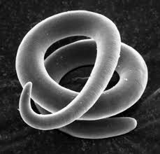
There are thousands of nematodes, or roundworms. Not only are there more than 15,000 known species of roundworms, but there are many thousands of individual nematodes in even a single handful of garden soil. Of the eighteen Orders in the phylum Nematoda, seven contain nematodes that are parasites or associates of invertebrates, and six include species that are parasites of vertebrate animals. The body of a nematode is long and narrow, resembling a tiny thread in many cases, and this is the origin of the group’s name. The word “nematode” comes from a Greek word nema that means “thread.” The “skin” of a nematode is highly unusual; it is not composed of cells like other animals, but instead is a mass of cellular material and nuclei without separate membranes. This epidermis secretes a thick outercuticle which is both tough and flexible. As other invertebrate groups, the cuticle is periodically shed during the life of a nematode as it grows, usually four times before reaching the adult stage.
Parasitic on Humans
Intestinal disease

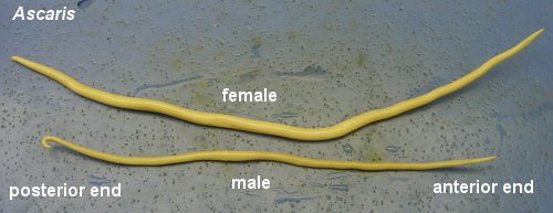
Ascaris lumbricoides is the largest nematode (roundworm) parasitizing the human intestine. (Adult females: 20 to 35 cm; adult male: 15 to 30 cm). People infected with Ascaris sp. often show no symptoms. If symptoms do occur they can be light and include abdominal discomfort. Heavy infections can cause intestinal blockage and impair growth in children. Other symptoms such as cough are due to migration of the worms through the body. Ascariasis is treatable with medication prescribed by your health care provider.
Pinworm Infection
Pinworm infection is caused by a small, thin, white roundworm called Enterobius vermicularis that can grow to be between 8-13 mm in the adult stage. Humans are considered the only hosts. Although pinworom infection can affect all people, it most commonly occurs among children, institutionalized persons, and household members of persons with pinworm infection. Pinworm infection is treatable with over-the-counter or prescription medication, but reinfection, which occurs easily, should be prevented.
Capillariasis
Capillariasis is a parasitic infection cause by two species of nematodes: Capillaria hepatica, which causes hepatic capillariasis, and Capillaria philippinensis, which causes intestinal capillariasis. C. hepatica is often found in the liver of animals such as small rodents, monkeys, and prairie dogs and can cause cirrhosis in these animal hosts. When these animals are eaten by larger carnivores, capillarid eggs are ingested and passed through the fecal matter of the carnivore. When this fecal matter is deposited in the soil, these eggs become infective in about 30 days and can infect other animals, including humans. Once accidentally ingested by a human, the eggs migrate to the liver and mature to adult worms. Another route of transmission is through the decomposition of infected animals via eggs in the liver being released into the soil.
C. philippinensis is often found in the tissues of small, freshwater fish. When humans ingest these raw or undercooked infected fish, larvae migrate to the intestine and mature to adult worms. Female worms deposit eggs in the intestine, which are released in fecal matter. When infected human fecal matter reaches freshwater, fish can become infected and the cycle continues. Some eggs hatch within the human intestine causing hyperinfection (a massive number of adult worms).
Life Cycle of Caillaria hepatica
Trichinosis

Trichinosis is a disease that people can get by eating raw or undercooked meat from animals infected with the microscopic parasite Trichinella. The first symptoms of trichinellosis are gastrointestinal, usually occurring 1-2 days after a person consumes raw or undercooked meat from a Trichinella-infected animal. These symptoms include nausea, diarrhea, vomiting, and abdominal pain. Late stage infection symptoms are muscle pain; fever; swelling of face, particularly eyes; weakness or fatigue, headache, chills, itchy skin or rash, cough, and some early stage symptoms too. Symptoms may range from very mild to severe and relate to the number of infectious worms consumed in the meat. If the infection is heavy, persons may have trouble coordinating movements, and have heart and breathing problems. Although rare, death can occur in severe cases. For mild to moderate infections, most symptoms go away within a few months.
Zoonotic hookworm infection
Zoonotic hookworms are hookworms that live in animals but can be transmitted to humans. Dogs and cats can become infected with several hookworm species, including Ancylostoma brazilense, A. caninum,A. ceylanicum, and Uncinaria stenocephala. The eggs of these parasites are shed in the feces of infected animals and can end up in the environment, contaminating the ground where the animal defecated. People become infected when the zoonotic hookworm larvae penetrate unprotected skin, especially when walking barefoot or sitting on contaminated soil or sand. This can result in a disease called cutaneous larva migrans (CLM), when the larvae migrate through the skin and cause inflammation. In rare cases, certain types of animal hookworm may infect the intestine and cause abdominal pain, discomfort, and diarrhea. Below is an image showing the progression of the hookworm infection.

Human hookworm infection
Hookworm is an intestinal parasite of humans. The larvae and adult worms live in the small intestine can cause intestinal disease. The two main species of hookworm infecting humans are Ancylostoma duodenaleand Necator americanus. Hookworm eggs are passed in the feces of an infected person. If an infected person defecates outside (near bushes, in a garden, or field) or if the feces from an infected person are used as fertilizer, eggs are deposited on soil. They can then mature and hatch, releasing larvae (immature worms). The larvae mature into a form that can penetrate the skin of humans. Hookworm infection is transmitted primarily by walking barefoot on contaminated soil. One kind of hookworm (Ancylostoma duodenale)can also be transmitted through the ingestion of larvae. People living in areas with warm and moist climates and where sanitation and hygiene are poor are at risk for hookworm infection if they walk barefoot or in other ways allow their skin to have direct contact with contaminated soil. Soil is contaminated by an infected person defecating outside or when human feces (“night soil”) are used as fertilizer. Children who play in contaminated soil may also be at risk.
Trichuriasis

Trichuriasis is an infection caused by whipworms (Trichuris trichiura), an intestinal parasite of humans. The larvae and adult worms live in the intestine of humans and can cause intestinal disease. The name is derived from the worm’s distinctive whip-like shape. Whipworms live in the intestine and whipworm eggs are passed in the feces of infected persons. If the infected person defecates outside (near bushes, in a garden, or field), or if the feces of an infected person are used as fertilizer, then eggs are deposited on the soil. They can then mature into a form that is infective. Roundworm infection is caused by ingesting eggs. This can happen when hands or fingers that have contaminated dirt on them are put in the mouth, or by consuming vegetables or fruits that have not been carefully cooked, washed or peeled. Infection occurs worldwide in warm and humid climates where sanitation and hygiene are poor, including in temperate climates during warmer months. Persons in these areas are at risk if soil contaminated with human feces enters their mouths or if they eat vegetables or fruits that have not been carefully washed, peeled or cooked.
Dirofilaria
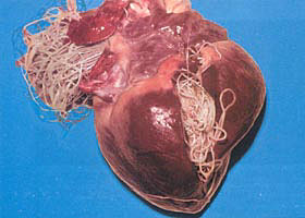
Dirofilaria is an infection of a various thin roundworms of the genus Dirofilaria and transmitted by mosquito bites. There are many species of Dirofilaria, but human infection is caused most commonly by three species, D. immitis, D. repens, and D. tenuis. The main natural hosts for these three species are dogs and wild canids, such as foxes and wolves (D. immitis and D. repens) and raccoons (D. tenuis). D. immitis is also known as “heartworm.” D. repens is not found in the United States, and D. tenuisappears to be restricted to raccoons in North America.
Dirofilariasis is the disease caused by Dirofilaria worm infections. In dogs, one form is called “heartworm disease” and is caused by D. immitis. D. immitis adult worms can cause pulmonary artery blockage in dogs, leading to an illness that can include cough, exhaustion upon exercise, fainting, coughing up blood, and severe weight loss.

In persons infected with D. immitis, dying worms in pulmonary artery branches can produce granulomas (small nodules formed by an inflammatory reaction), a condition called “pulmonary dirofilariasis.” The granulomas appear as coin lesions (small, round abnormalities) on chest x-rays. Most persons with pulmonary dirofilariasis have no symptoms. People with symptoms may experience cough (including coughing up blood), chest pain, fever, and pleural effusion (excess fluid between the tissues that line the lungs and the chest cavity). Coin lesions on chest x-rays are not diagnostically specific for pulmonary dirofilariasis. Therefore, discovery of these lesions have led to invasive diagnostic procedures to exclude other, more serious causes, including cancer. Rarely, D. immitis worms have been found in humans outside the lungs, including in the brain, eye, and testicle.