Nutritional Adaptations in Animals -working copy
Lab Information- Please read the entire Lab Unit 9 and work through all activities and answer questions in your lab manual using the lab text and website. Read and follow this outline modification for this lab.
BLENDED –FIND TERMS THAT WERE OMITTED? LOOK IN UNIT 9? CAN’T FIND IN WORDPRESS.
What do termites and cows have in common? Remember looking at these during the Species Relationship lab?
– proposed F2020
insert pre-lab assignment handout]
- Activity #1: Paramecium Feeding (online videos) p. 153-155. Complete steps #5 & 8 in your lab manual.
- Activity #2: Hydra Feeding (online videos) p. 154-155 complete step #4 in your lab manual.
- Activity #3: Bullfrog Dissection (online videos) p. 155-157. Use one or more of the online videos or animations to complete the information in the lab manual (steps #7-11).
- Read the background information on Mammals’ Teeth (p. 158-160) for use IN-LAB.
Feeding Strategies
Understand the terminology associated with the manner of Food Acquisition and Digestion, and Absorption of Nutrient strategies (see bold terms in your lab manual)
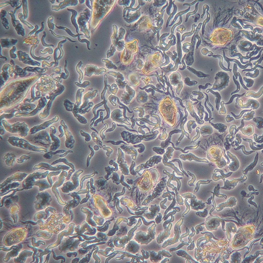
Activity One: Paramecium Feeding (p. 153-154)
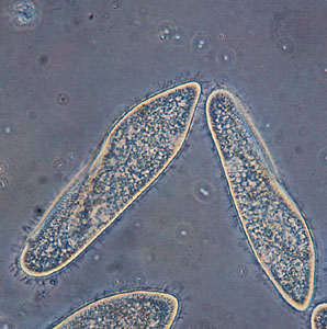
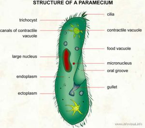
compound light microscope (at 400X) and diagram (left and right image respectively)
- Description: ciliate Protozoan
- Habitat: stagnant freshwater
- Movement: cilia
- Nutrition: An oral groove is used to ingest nutrients and food vacuoles are responsible for digestion.
- Distinct Layers: The cytoplasm of protozoans can be divided into 2 layers. The ectoplasm is the more clear outermost layer and the endoplasm is the central area surrounding all organelles.
Videos of Paramecium feeding (#1 is what you would have seen in lab)
- Paramecium eating Congo Red stained yeast cells. As the yeast cells enter the Paramecium, they are encapsulated into food vacuoles. When the yeast cells get digested by acidic enzymes, the red stain will change the yeast cells from red to blue.
- At higher magnification you can see the movement of the cilia around the perimeter of the cell used for movement, but also along the oral grove into the gullet.
- Closer still you can observe the cilia along the oral grove and the movement of food particles flowing into the cell. A food vacuole forms as particles get encapsulated to form a vacuole, which are then is released into the cytoplasm of the cell.
- After observing this feeding behavior, what terms can you apply to this type of feeding (Food Acquisition) and Digestion and Absorption of nutrients?
Activity Two: Hydra Feeding (p. 154-155)
- What key characteristics can you see above that helps you know that Hydra are part of the phylum Cnidaria?
- How do Hydra get food, ingest it, digest it, get the nutrients, and get rid of waste? -use the proper terms for these processes.
- Watch as a tiny Hydra rips its mouth open to feed. (Here is a short article that further explains how this works).
Videos of Hydra feeding (#2 is similar to what you would have seen in lab feeding the hydra Daphnia or brine shrimp)
- Video of Hydra eating a mosquito larva. Note the overall structure of the Hydra and the smaller bulb-like stinging cells (nematocysts) along the tentacles. Though the mosquito larva is quite large, the lack of a skeleton and jelly like body allows the Hydra a lot of stretching to fit food items through the mouth and into the gastrovascular cavity for digestion. How does the waste then get out?
- Green Hydra eating 3 water fleas (Cyclops). The Green Hydra have green algal endosymbionts that provide some carbohydrates to the Hydra, but not so much that they still need to eat small zooplankton.
- Describe how Hydra get food, ingest it, digest it, get the nutrients, and get rid of waste?
- After observing this feeding behavior, what terms can you apply to to the Hydra in for feeding (Food Acquisition) and Digestion and Absorption of nutrients?
Activity Three: Bullfrog Dissection (p. 155-157)
Images of Frog Dissections
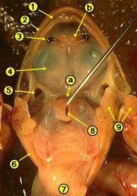 |
Frog Oral Cavity
a. Esophagus opening b. Vomerine teeth 1. Groove of the maxilla 2. Maxillary teeth 3. Internal nare 4. Eye ball 5. Eustachian tube opening 6. Jaw ridge 7. Underside of the tongue 8. Glottis 9. Cut muscle and bone
|
 |
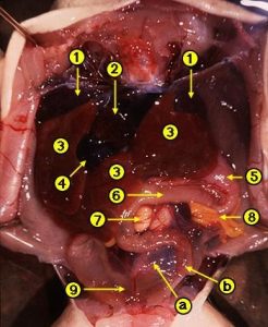
Internal – Female Frog
1. Liver lobes 2. Heart 3. Ovary with eggs 4. Gall bladder 5. Small intestine 6. Stomach 7. Oviduct
|
 |
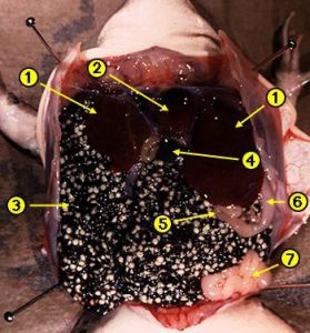
Internal – Male Frog
a. Large intestine b. Small intestine 1. Lung lobes 2. Heart 3. Liver lobes 4. Gall bladder 5. Stomach 6. Small intestine 7. Testis 8.Fat Body 9. Urinary bladder |
Videos of A Preserved Bullfrog dissection
- Review of external anatomy and internal mouth structures. This video nicely shows the interesting attachment of the tongue and its function in capturing prey. You will also see the 2 types of teeth used to help hold prey: the maxillary and vomerine teeth.
- Review of Internal anatomy of the digestive, and the circulatory, respiratory and excretory systems.
- These dissections do not show the reproductive structures in the male and female bullfrogs. For those refer to the images above or look at the animation below for the reproductive system.
Non-Dissection Alternatives:
Good Internal and External Anatomy with narration – (requires Flash Player, click to enable) – the initial cut and Digestive system show images of an actual preserved frog dissection (not animation) Other Internal Anatomy images are animated.
Good written summaries and pictures of important structures and dissection. The discussion of the mouth structures explain how the tongue is used
to capture prey and points out the 2 types of teeth (maxillary and vomerine teeth).
[enhanced-heading heading=”IN-LAB” headingtype=”h5″ headingstyle=”h2″ target=”_top” icon=”ucomm-microscope” iconstyle=”circle-filled” iconposition=”stacked” coloroptions=”text” textcolor=”wolfpackred” fontface=”universlight” /]
?Working in breakout rooms?
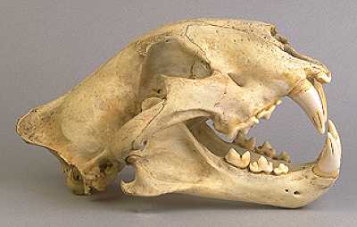
- TA leads discussion on Dental formulas, as a group, complete Table 9-1 Human Dentition.
- Review the provided information on Skull Structure and Muscles of Mastication.
- Complete pages 161-164 using the provided Mammal Skulls & Bird Beaks and Skulls.
- ??Could do 1 or 2 of the skulls together and the rest as POST-LAB assignment individual ???
- OR do as a group if also working on turning in final group projects???
Mammal Skulls and Dentition:
Cool skull website – California Academy of Sciences – click on “Enter the 3D gallery”
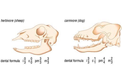
Activity Four: Mammals’ Teeth and Bird Bills (p. 158-166) -from Sp2020
This activity will allow you to examine mammal and bird skulls to gain knowledge about feeding and life style behaviors in a variety of animals. We will pay particular attention to examining the dentition, determining dental formulae, and looking for clues in Overall Skull Structures (see link below). Read the background information in the lab manual and use the image sets below to fill in Tables 9-1, 9-2, 9-3, 9-4 for 6 different mammals and Table 9-5 for the four birds. You will need to fill in all these charts and then send images of your completed charts to your TA as part of the 10 pt lab assignment for this week.
Skull Structures and Muscles of Mastication – Additional Information to help interpret feeding habit clues seen in skulls beyond looking at teeth.
View Mammals Skulls
View Bird Beaks and Bills
Birds have various beak sizes and shapes for a variety of reasons. However, the most important factor in determining beak shape and size is feeding habits.
Explore the various types of Bird Beaks and Bills!
[enhanced-heading heading=”POST-LAB” headingtype=”h5″ headingstyle=”h2″ target=”_top” icon=”ucomm-microscope” iconstyle=”circle-filled” iconposition=”stacked” coloroptions=”text” textcolor=”wolfpackred” fontface=”universlight” /]
???????????
[callout bodyfontface=”universroman” heading=”Nutritional Adaptation Assignment” headingicon=”noicon” textalign=”textcenter” url=”https://wordpress-projects.wolfware.ncsu.edu/bio-181l-zchxzbn/wp-content/uploads/sites/75/2020/03/Nutrition-Assignment-Sp2020.docx” type=”basic” bgcolor=”huntyellow” margin=”normal”]Use the attached Word guidelines/template to create ONE document to turn into your TA via Moodle as a PDF. This is due April 6-10 by midnight/11:59PM on the day of your regular lab section. You will work your way through all the activities and questions in the Nutrition lab using the lab manual and the lab website. This assignment is worth 10 points.[/callout]