Diversity in Transport (a 2 Week lab unit) – Online –working copy??
?Change format, Pre-Lab, In-Lab, Post Lab
[enhanced-heading heading=”PRE-LAB” headingtype=”h5″ headingstyle=”h2″ target=”_top” icon=”ucomm-heart” iconstyle=”circle-filled” iconposition=”stacked” coloroptions=”text” textcolor=”wolfpackred” fontface=”universlight” /]
[insert pre-lab assignment handout]
- Activity 1: Transpiration in Higher Plants (p. 168-169).
- Focus on Plant Anatomy and Transpiration/Translocation – substitute the potometer procedure with Simulation from Glencoe (requires FlashPlayer) OR try Pratt simulation to examine many factors – PS, Cell Respiration, Absorption, Transpirations and Translocations. -make an assignment for this? Not sure what Erica did with this.?? No longer graphing data, but looking more holistically???
- Activity 2: Earthworm Dissection (p. 175-176)
- Activity 3: Crayfish Dissection (p. 176-179.
I will reorganize/move the content below once I know what is Pre-Lab, In-Lab, Post-Lab AND if 1 or 2 weeks of Transport.
Work on Week 2 IN-LAB ???? if we do this all in one week?
- Focus on Heart?
[enhanced-heading heading=”POST-LAB” headingtype=”h5″ headingstyle=”h2″ target=”_top” icon=”ucomm-heart” iconstyle=”circle-filled” iconposition=”stacked” coloroptions=”text” textcolor=”wolfpackred” fontface=”universlight” /]
I will reorganize the content below once I know what is Pre-Lab, In-Lab, Post-Lab AND if 1 or 2 weeks of Transport.
Lab Information- Please read the Lab Unit 10 week 1 (pages 167-182), work through all activities and answer questions in your lab manual using the lab text and website. Read and follow this outline modification for the Transport lab week.
Transport in Plants: (Week 1)
Background:
- What is transported?
- Which tissues (xylem and phloem) are used for Transpiration? for Translocation?
- What is the difference between Transpiration and Translocation?
- What drives these types of transport?
- Leaf Anatomy (though stomates are not generally found on the upper epidermis as seen here, this diagram nicely shows important cell layers and gas exchange)
- Stomata opening and closing – videoclip (1:07)
- Water Transport in Plants – short video David Attenborough (3:05) -video continues to 4:44, but not as applicable to our experiment.
- Plant Transport: Xylem, Phloem and Transpiration (3D Animation) (5:36) More detailed than needed for lab, but very good and may be helpful for understanding the background if not yet covered in lecture.
We will look more closely at structures involved in both Transpiration and Translocation in plant leaves and stem cross sections.
Text and Figures in the Lab Manual pages 95-96 and 169 will be very helpful for interpreting and labeling your drawings of the following slides.
Use the empty space on page 180 to draw and label the pictures #1, #2, #3, and #4 below – Sp2020.
#1. 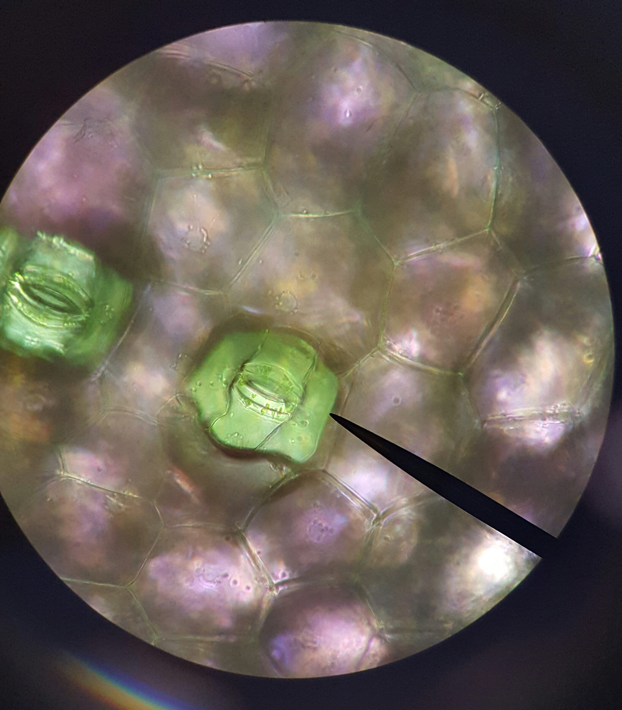 . #2.
. #2. 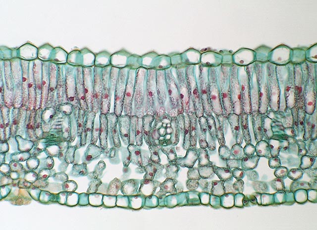
Draw and label the following prepared slides of plant tissues. – Sp2020
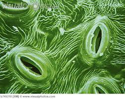
- Picture #1 is a view of the lower epidermis of Zebrina . It is from a whole mount or epidermal peel preparation.
- Make a fresh epidermal leaf peels of a Zebrina leaf to study stomate structure.
- Peel the leaf to expose the lower epidermis without other cell layers. If you cannot get a good leaf peel, then make a wet mount of a thin area of the leaf.
- Place the peel or leaf piece epidermis side up (more purple side) facing UP on the slide. Then make a wet mount (top with a drop of water and a coverslip). View with the compound microscope – medium to high power.
- Students should sketch what they see on the lower epidermis. Structures to draw and label: epidermal cells, guard cells, stoma (= singular) or stomata/stomates (= plural).
- Leaf Cross section slide (#2) – Fig. 6-2, p. 95 and Fig. 10-2 p. 169 – Structures to draw and label: cuticle, upper and lower epidermis, guard cells (stoma), mesophyll (contains spongy mesophyll and palisade layers), vein or midvein containing xylem and phloem.
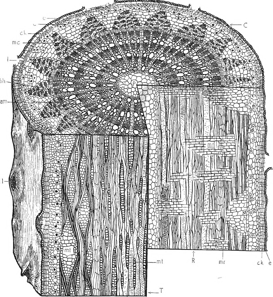
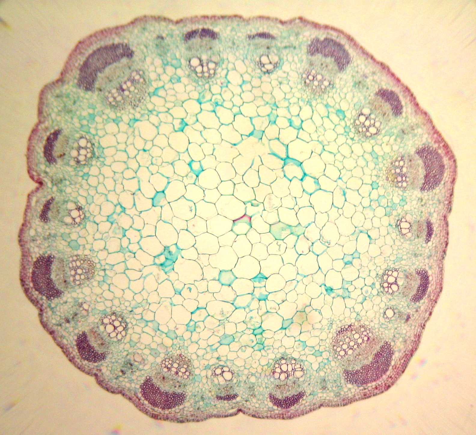
- Herbaceous (#3) and Woody Stem slide (#4) – Fig. 6-4, p. 96 and Fig. 10-1, p. 169 – Epidermis, cortex, pith, vascular bundles in herbaceous stems (xylem/inside and phloem/towards outside of bundles), in woody stem xylem collects as inner stem annual rings (eventually growth rings/wood), phloem to the outside under the cork (in woody stems), epidermis layer.
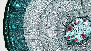
- Tilia (# 4) woody stem sections showing 1, 2, 3 & 4 growth rings – Same woody stem as the slide in #3 above) – Fig. 6-4A, p. 96 – should be able to see most of the same structures as the woody stem above, but more developed in the older stem sections –pith is smaller on the inside, more developed annual rings/xylem accumulation with age that becomes wood, phloem to the outside of the xylem rings being crushed under the cork/epidermis layers, cortex less and less apparent under outmost layers.
Spring 2020: Due to the change to online labs, we will not be able to do Activity One in the lab manual. Instead we will use the following simulation.
Simulation of Plant Transpiration Experiment – you may need to enable Flash Player.
- Follow the directions as you read and scroll down using the information on the left side in the simulation.
- Work through the simulation selecting all 4 plants and working through the control and 3 different treatments for each. Collect the data after each “run”.
- Use the data to make ONE graph including data from all 4 plants under control and all treatment/environmental conditions. Be sure to include all of the appropriate components of a proper graph (see Appendix D Excel in the lab manual).
Cool Articles/links About Researchers Using Plants for Phytoremediation
- Researchers in Elizabeth City NC – ‘Green Clean:’ Researchers Determining Natural Ways to Clean Contaminated Soil
- The Effects of Urban Trees on Air Quality (short article)
- NCSU Master of Environmental Assessment Degree Program
Transport in Animals: (Week 1)
Open and Closed Circulatory Systems
Activity 3 (p. 177-179) Crayfish – open circulatory system
- Open circulatory system – The open circulatory system is common to invertebrates. In an open circulatory system, the blood is pumped into a hemocoel and diffuses back to the circulatory system between cells. Blood is pumped into body cavities by a heart. See hemolymph circulation in Insects (0:48), discussion of circulation in a cockroach (1:45).
Crayfish Dissection Images
- External Anatomy: Anterior
- External Anatomy: Posterior
- External Anatomy: Mouthparts
- Internal Anatomy: Lateral
- Internal Anatomy: Dorsal Anterior
- Female Crayfish: Ventral
- Male Crayfish: Ventral
Videos of Preserved Crayfish Dissections
- Crayfish Dissections – External Anatomy (8:52) and Internal Anatomy (5:55)
- Crayfish Dissection – Includes both the external and internal anatomy (7:14)
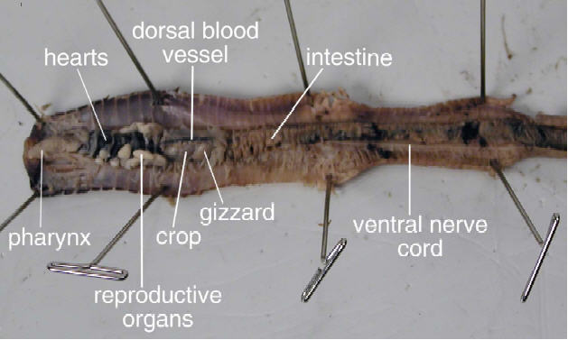
Activity 2 (p. 175-176) Earthworm – closed circulatory system
- Closed circulatory system – Vertebrates, and some invertebrates, have a closed circulatory system. In a closed circulatory system, the blood is in closed in a vessel at all times. The blood is pumped by a heart and travels through each of the closed vessels. The blood is not typically pumped into any body cavities.
Earthworm
Note the closed circulatory system
Earthworm Dissection Animation – You may need to enable Flash Player.
You will need to take screen shots of your fully labeled External and Internal Earthworm Anatomy to submit with your assignment.
- Label the indicated structures for external anatomy in this animation.
- Label the indicated structures for internal anatomy in this animation.
Videos of Preserved Earthworm dissections
- Preserved Earthworm Dissection Video – external and internal anatomy (7:27)
- Earthworm Dissection video (8:49)
[callout heading=”Transport Week 1 Assignment” headingicon=”noicon” textalign=”textcenter” url=”https://wordpress-projects.wolfware.ncsu.edu/bio-181l-zchxzbn/wp-content/uploads/sites/75/2020/04/BIO-181-Transport-1-Assignment-Sp2020.docx” target=”true” type=”basic” bgcolor=”huntyellow”]Use the attached Word guidelines/template to create ONE document to turn into your TA via Moodle as a PDF. This is due April 13-17 by midnight/11:59PM on the day of your regular lab section. You will work your way through all the activities and questions in the Transport Week 1 lab using the lab manual and the lab website. This assignment is worth 10 points.[/callout]
???I could make this a separate page OR add in Pre, In and Post lab icons here. I think these were kept as ONE page since the heart info spans 2 lab units.
Transport in Animals: (Week 2)
Lab Information- Please read the Lab Unit 10 week 2 (pages 183-195), work through all activities and answer questions in your lab manual using the lab text and website. Read and follow this outline modification for the Transport lab week.
You will label drawings of plant structures, perform a Transpiration simulation, graph your data and answer questions that you will submit to your TA in Moodle.
???*If you do not have your lab manual, you can print out the indicated pages (questions and diagrams) or write all your work out on paper to save and use for studying for your Unit 4 Quiz/Assignment that will be given in the lecture Moodle class April 23 (sect. 001) and 24 (sect. 002) lecture sections.
Mammalian Circulation
We will focus this week of lab on:
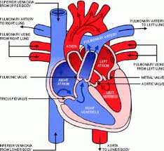
- the 4 chambers of the heart
- right vs. left
- where did blood come from when entering the heart? -> right atria, -> left atria?
- where does it go when it leaves the heart? -> right ventricle, -> left ventricle?
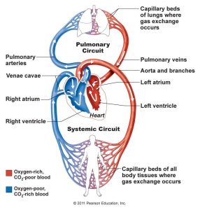
- where is the blood oxygenated and deoxygenated
- pulmonary vs. systemic circulation
- interpreting EKGs (what occurs at the PQRST peaks).
- how does activity affect heart rate (BPM) and why?
Activity One: Heart Structure and Double Circulation (Pulmonary and Systemic)
Animation – Heart – Systole and Diastole
Video – External Anatomy of the Heart and Blood Flow (using a heart model)
Video – Internal Anatomy of the Heart and Blood Flow (using a heart model)
Mammalian Heart – Anatomy and Function – helpful links
- Label a heart
- View pulmonary circulation
- Human Circulatory System (images from Human Anatomy, 2nd Ed. McKinley and O’Loughlin)
- Circulation Rap/Song
- Sheep heart dissection – similar to what you would have seen in lab
- Living with an Artificial Heart: Prince’s Journey (5:45)
- Could artificial hearts make donations obsolete? (4:16)
Activity Two: Heart Function and ECGs
Electrocardiogram – nice background information on electrical activity in the heart (1:20)
ECG peaks and heartbeats
ECG – Example of a student ECG from our lab (recording over 10 seconds)
EKG Online Course Introduction – Check out the lessons found in the Table of Contents and the EKG Basics Quiz at the bottom of the page. This is not to be used for self diagnosis or diagnosing others.